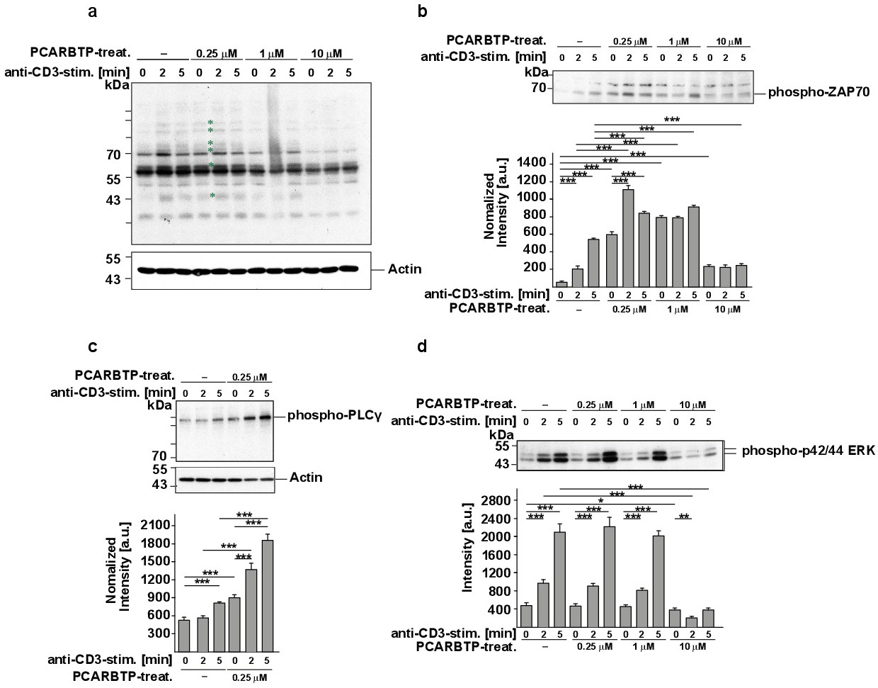
Fig. 1. Mitochondria-targeted Kv1.3 inhibitors induce basal tyrosine phosphorylation of several cellular proteins including ZAP70 and PLCg. Jurkat cells were left untreated or treated with 0.25 mM, 1 mM or 10 mM PCARBTP in the presence or absence of the anti-CD3 antibody OKT-3 (1 mg/mL) for the indicated times. Cells were lysed, insoluble material was removed, the samples were separated on 7.5 - 10% SDS-PAGE and blotted for cellular tyrosine phosphorylation (a), phospho-ZAP70 (b), phospho-PLCg (c) or phospho-ERK (d). Asterisks in panel a indicate proteins that show an increased tyrosine phosphorylation after treatment with low doses of PCARBTP. Blots determining phospho-ZAP70, phospho-PLCg or phospho-ERK were quantified using Image J. The quantification is shown in the bar graphs in each Fig.. Panel d shows the quantification of p42, the results for p44 are very similar (not shown). The results show a constitutive phosphorylation/activation of several cellular proteins including ZAP70 and PLCg after treatment with low doses of PCARBTP. (c) Neither basal nor OKT-3-induced phosphorylation of ERK is altered by treatment with low doses of PCARBTP. High concentrations (10 mM) of PCARBTP reduce basal and OKT-3 induced cellular tyrosine phosphorylation. Aliquots of the samples were blotted for actin to show that similar amounts of proteins were loaded in all gels. Panel a, c, and d were performed from the same aliquots and the corresponding actin blot is displayed in panel a. Shown are representative blots from 4 independent studies or the mean ± SD, n=4, *p<0.05, **p<0.01, ***p<0.001, ANOVA.